2 Spots On Lung
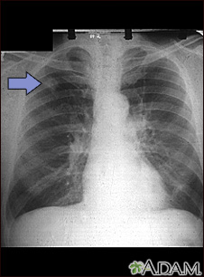
Nodules or spots in the lungs may be due to a variety of issues such as infection including conditions such as histoplasmosis, sarcoidosis, and pneumonia. Additional diagnostic tests may need to be done. To ease any anxiety and for proper evaluation, it is best that you discuss this with your attending physician also. Ground glass opacities, referring to findings on computed tomography (CT) scans of COVID-19 patients, can diagnose coronavirus infections—but what exactly are 'ground glass opacities' in lung scans? A lung cancer goes through 40 doublings of volume in its life cycle and is not visible until it reaches 1 cm in size, Dr. At 3 to 4 cm, it has gone through seven eighths of its life history. 'By the time it reaches 2 cm, the tumor contains 500 million malignant cells and starts to metastasize,' he said. A spot on the lungs usually refers to a pulmonary nodule. This is a small, round growth on the lungs that shows up as a white spot on image scans. Typically, these nodules are smaller than three 3.
New Patient Appointment
or214-645-8300
New Patient Appointment or214-645-8300
Lung nodules are soft-tissue lesions that can be either rounded or irregular in shape. A nodule is defined as a lesion measuring 3 centimeters or smaller in diameter, says lung specialist Louis.
New Patient Appointment or214-645-8300
A solitary pulmonary nodule or “spot on the lung” is defined as a discrete, well-defined, rounded opacity less than or equal to 3 cm (1.5 inches) in diameter that is completely surrounded by lung tissue, does not touch the root of the lung or mediastinum, and is not associated with enlarged lymph nodes, collapsed lung, or pleural effusion.
A pulmonary nodule can be benign or cancerous. Lesions larger than 3 cm are considered masses and are treated as cancerous until proven otherwise.
Lung nodules are quite common and are found on one in 500 chest X-rays and one in 100 CT scans of the chest. Lung nodules are being recognized more frequently with the wider application of CT screening for lung cancer. Roughly half of people who smoke over the age of 50 will have nodules on a CT scan of their chest.
The thoracic surgeons and interventional pulmonologists at UT Southwestern Medical Center perform leading-edge procedures to evaluate and treat pulmonary nodules and various lung lesions – including bronchoscopic procedures, image-guided sampling, conventional surgical procedures, and more advanced minimally invasive and robotic techniques.
We feature the latest imaging techniques and treatments through our advanced imaging center, including endobronchial ultrasound (EBUS), electromagnetic navigational bronchoscopy (ENB), and many other techniques.
Our surgeons work closely with UT Southwestern’s interventional pulmonary team, oncologists, radiologists, and pathologists to deliver comprehensive care – all in one location, and usually on the same day. The pulmonary nodule clinic at UT Southwestern streamlines the evaluation and management of patients with pulmonary nodules and abnormal findings on imaging, including screening CT scan.
Causes of Lung Nodules
Lung nodules can be either benign (noncancerous) or malignant (cancer). The most common causes of benign nodules include granulomas (clumps of inflamed tissue) and hamartomas (benign lung tumors).
The most common cause of cancerous or malignant lung nodules includes lung cancer or cancer from other regions of the body that has spread to the lungs (metastatic cancer).
Lung nodules can be divided into a few major categories:
- Benign tumors, such as hamartomas
- Infections, including bacterial infections such as tuberculosis, and fungal infections such as histoplasmosis and coccidiomycosis
- Inflammation, such as rheumatoid arthritis, sarcoidosis, and Wegener's granulomatosis
- Malignant tumors, including lung cancer and cancer that has spread to the lungs from other parts of the body.
Overall, the likelihood that a lung nodule is a cancer is approximately 40 percent, but the risk of a lung nodule being cancerous varies considerably depending on several things, including:
- Age: Rare in people under 35 years of age. Half of lung nodules in people over age 50 are malignant
- Calcification: Lung nodules that are calcified are more likely to be benign
- Cavitation: Nodules described as “cavitary,” meaning that the interior part of the nodule appears to contain air on X-rays, are more likely to be benign
- Growth: Cancerous lung nodules tend to grow fairly rapidly with an average doubling time of about four months, while benign nodules tend to remain the same size over time
- Medical history: Having a history of cancer increases the chance that it could be malignant
- Occupation: Some occupational exposures raise the likelihood that a nodule is a cancer
- Smoking: Current and former smokers are more likely to have cancerous lung nodules than never smokers
- Size: Larger nodules are more likely to be cancerous than smaller ones
- Shape: Smooth, round nodules are more likely to be benign, while irregular or “spiculated” nodules are more likely to be cancerous
Diagnosis
If we suspect that you have a pulmonary nodule, we will conduct a physical examination and order tests to confirm the diagnosis. Studies to evaluate and diagnose pulmonary nodules might include:
- Bronchoscopy, including advanced guided techniques such as endobronchial ultrasound (EBUS), electromagnetic navigational bronchoscopy (ENB), and other procedures
- Chest X-rays (radiographs)
- Computed tomography (CT), including screening CT scan
- Fluoroscopy: Real-time X-ray imaging
- Image-guided sampling techniques that include CT-guided and ultrasound-guided biopsies (fine needle aspiration biopsy or FNA)
- Magnetic resonance imaging (MRI)
- Minimally invasive lung biopsy (thoracoscopic or robotic)
- Positron emission tomography (PET)
- Pulmonary function studies (PFT)
Based on the characteristics and size of the lung nodule on the CT scan, we may recommend:
- Observation and repeat X-ray studies if the nodule is likely benign
- Further imaging, such as a repeat CT scan of your chest or a PET scan
- Biopsy of the nodule via bronchoscopy (if the nodule is near one of your airways), a needle biopsy (if the nodule is located near the outside of your lungs), or lung surgery (video-assisted thoracoscopic surgery) if we think it may be malignant
- Treatments
When surgery is the most appropriate therapy, our thoracic surgeons treat pulmonary nodules and lung lesions with procedures that include:
- Lobectomy: Removal of an entire lobe by minimally invasive VATS or by robotic techniques
- Segmentectomy: Removal of a segment of a lobe by minimally invasive video-assisted thoracoscopic surgery (VATS) or by robotic techniques
- Wedge resection: Removal of the lung nodule along with a small amount of lung tissue
Minimally Invasive Surgery
Compared to surgery performed through an open chest incision, minimally invasive surgery provides several important benefits for patients, including:
- Faster recovery and return to normal activities
- Shorter hospital stay
- Less pain
- Little scarring
- Minimal blood loss
- No cutting of the ribs or sternum
When surgery is not possible in a patient with a cancerous or malignant nodule, our multidisciplinary team of surgeons and medical and radiation oncologists will provide recommendations about the best management options. These options may include advanced radiation techniques, systemic therapy with conventional chemotherapy, and/or targeted or personalized therapies.
Clinical Trials
UT Southwestern conducts clinical trials aimed at improving the treatment of pulmonary nodules. Talk with your doctor to see if a clinical trial may be right for you.
Related Conditions and Treatments
Find a Clinical Trial
Search for opportunities to participate in a lung disease or asthma research study.
Award-Winning Care
We’re one of the world’s top academic medical centers, with a unique legacy of innovation in patient care and scientific discovery.
MyChart
Our secure online portal for patients makes it easy to communicate with your doctor, access test results, and more.
Related Clinics
Showing 5 locations
Professional Office Building 2
5939 Harry Hines Blvd.Dallas, Texas 75390 214-645-8300
Thoracic Surgery
at UT Southwestern Harold C. Simmons Comprehensive Cancer Center at Moncrief Cancer Institute 400 W. Magnolia AvenueFort Worth, Texas 76104 214-645-7700Directions
University Hospital Medical Oncology Clinic - Pulmonary
at Seay Biomedical Building 2201 Inwood Road, 3rd Floor, Suite 500Dallas, Texas 75390 214-645-4673
University Hospital Outpatient Imaging Services - CT Lung Screening
at Outpatient Building 1801 Inwood Road, 1st FloorDallas, Texas 75390 214-645-3453
University Hospital Simmons Comprehensive Cancer Clinic
at UT Southwestern Medical Center at Richardson/Plano 3030 Waterview Parkway, 2nd FloorRichardson, Texas 75080 972-669-7077
 Directions
 2 Spots On Lung
I have been spotted with a spot in the lung: what can it mean? If you have been having symptoms such as cough, expectoration with blood and/or chest pain when breathing, it is likely that you have already seen the doctor, have requested a chest x-ray and have detected spots in the lungs.
First of all you should know that not all the spots in the lungs are blood. It is normal that you feel worried about this situation and, although cancer is the cause of most concern, most of the time these lesions or spots appear, they are due to other reasons. In the following article we will talk about the spots in the lungs: causes and symptoms.
What it means when there is a spot on the lung
Not all the spots in the lungs are alarming; infections are actually the most common causes of lung injury. Among the most common infections in which there is evidence of lung spots are:
Pneumonia: An infectious disease that occurs in one or both lungs, usually of bacterial origin. The injuries on the radiography represent the congested alveoli of secretions. The image shows an irregular white spot, commonly located at the base of the lung.
Tuberculosis: Chronic and progressive infection, frequent in people with a weakened immune system such as HIV. Tuberculosis can cause spots in the whitish lungs observable in the X-ray. It also presents with weight loss and cough with expectoration of more than 15 days of evolution, characteristic symptomatology.
Granuloma: There are nothing more than masses of immune cells formed as a mechanism of self-defense when the body is faced with foreign substances. There are diseases that are characterized by being granulomatous, for example: tuberculosis, histoplasmosis and coccidioidomycosis.
Lung stain on the CT scan due to pulmonary edema
The pulmonary edema is medically defined as the accumulation of fluid inside the lungs. Among the common symptoms of pulmonary edema are:
- Expectoration with blood
- Pallor
- Excessive Sweating
- Anxiety
- Tachycardia
This pathology represents a medical emergency, so if you notice these symptoms you should visit the doctor. Pulmonary edema represents the second cause of lesions in the lungs when a doctor evaluates a chest x-ray, they are also seen as lightly blurred spots in the pulmonary surroundings, the frequent symptom of this disease is shortness of breath or dyspnea.
At the time of visualizing a lesion on the lungs, should not be overlooked pulmonary fibrosis in which the lung heals and therefore this tissue will not receive adequate oxygen. Although its causes are unknown, smoking tobacco is the most common cause.
A spot on the lung: what can it mean?
2 Spots On Lung
If you have detected a spot in the lung you should know that there are other pathologies that can also be detected through chest x-rays, these spots can be cancerous and non-cancerous growths. Among those that are not cancerous, we find:
Hemartoma: These are disorganized tissues that can reach large sizes similar to tumors but called malformation. They can develop anywhere in the body standing out in the lungs.
2 Mm Spot On Lung
Lipoma: These are benign fatty tissue tumors, are usually localized in the skin but can appear anywhere or organ with fatty tissue, where the lungs are included. The risk factor for the appearance of pulmonary lipoma highlights tobacco smoke. It is evident in the X-ray as a well-defined rounded mass.
On the other hand, lung cancer is the most serious reason related to lung spots, it is frequent in heavy smokers, and in fact it turns out to be the disease with the highest mortality worldwide. The most dangerous aspect of this disease is that its course is asymptomatic or silent, however, when it is diagnosed, it already has characteristic advanced symptoms. The chest radiography shows nodules of small or large size, depending on the stage of lung cancer, which are observed as a white spot on the radiography.
Symptoms of lung spots
Determine the spots in the lungs can arise by chance by routine studies or presenting the common symptoms of respiratory diseases such as:
- Cough with expectoration.
- Hemoptysis.
- Dyspnea or shortness of breath or difficulty to repair.
- Pain in the chest when breathing.
- Dysphonia or hoarseness.
- Night sweats.
- Weightloss.
- Fever.
Detected a spot on my lung: What should I do?
2 Spots On Lungs
The lungs are fundamental for life. The ideal is to prevent the appearance of some spot or lung injury with good habits, however, these tips can also serve to keep the lungs healthy:
2 Spots On Lungs
- By exercising to have resistant lungs, walking or swimming is recommended.
- Quitting smoking, most of the lung diseases where the spots visualized in the TACs are prevalent are caused by tobacco.
- Reducing fat consumption by allowing clean lungs.
- It is also recommended to detoxify the lungs for 3 days based on fruits and vegetables and ingesting plenty of water, decreasing the consumption of coffee sugar and processed foods.
- The use of the carrot strengthens the mucous membranes of the lung.
- On the other hand, radish is a natural antibiotic can be used in infusions and salads.
- Garlic is a natural product that allows you to breathe better by eating 1 garlic on an empty stomach daily.
- It is recommended to use valuations with eucalyptus to clean the respiratory system.
- Practice breathing exercises, vital to enjoy healthy lung. Inspiring slowly across the nose counting up to 5 mentally and exhaling for the mouth at the same time. Repeat this exercise 5 times a day.
2 Inch Spot On Lung
Obviously the causes are multiple but the main trigger is smoking, however, it is important that before being alarmed by the spots in the lungs go to a pulmonologist specialist who will request the complementary test to determine the proper diagnosis and the treatment you need.